
What is Keratoconus?
Your cornea is the clear, dome-shaped surface at the front of your eye that focuses light into the eye. Keratoconus is when the cornea progressively thins and bulges outward, forming a cone shape.
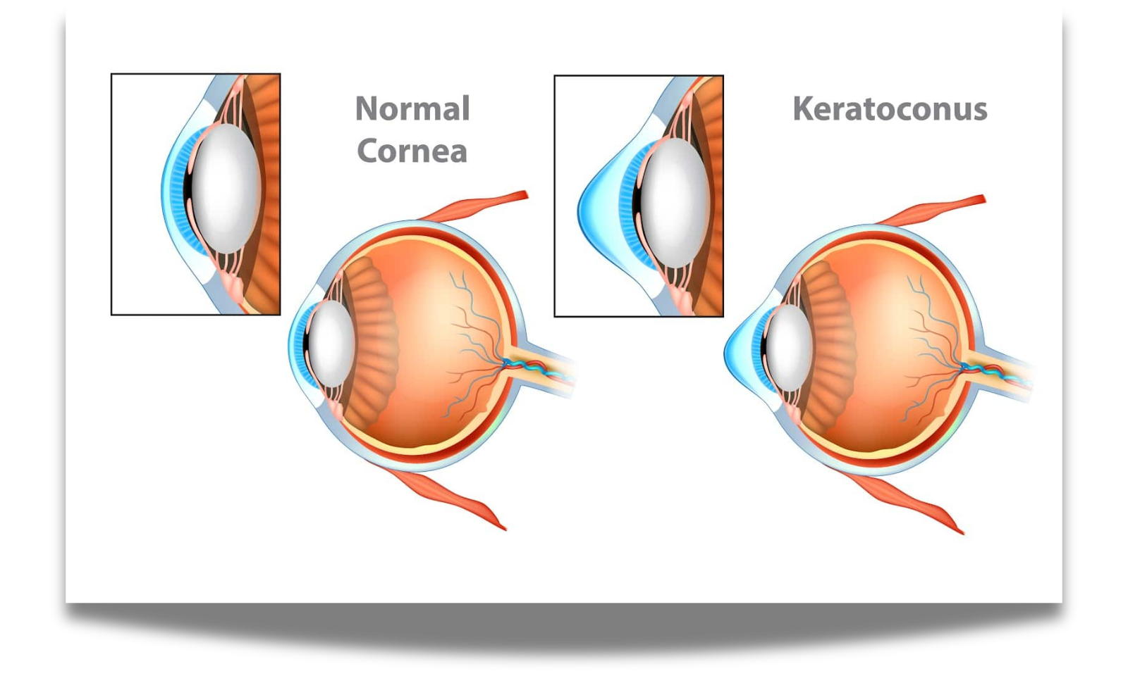
Keratoconus Symptoms
Keratoconus often affects both eyes, and symptoms can often differ in each eye.
In the early stage, symptoms can include: mild blurring of vision, slightly distorted vision, increased sensitivity to light and glare, eye redness or swelling.
In later stages, symptoms often include: more blurry and distorted vision, increased nearsightedness or astigmatism, not being able to wear traditional contact lenses.
Signs of keratoconus typically begin when people are in their late teens to early 20s and can advance into a person’s 30s. Keratoconus usually takes years to progress from early to late stage, although some individuals can get worse quickly.
Keratoconus Diagnosis
Keratoconus can be diagnosed through a comprehensive eye exam. During this exam we will take specialized measurements of your cornea to examine it’s curvature and the condition of the cornea’s surface. Annual monitoring is strongly recommended to detect early changes.
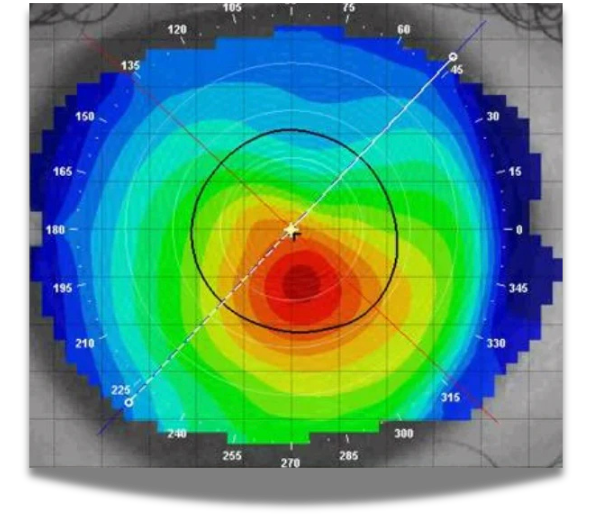
Corneal Topography showing Keratoconus
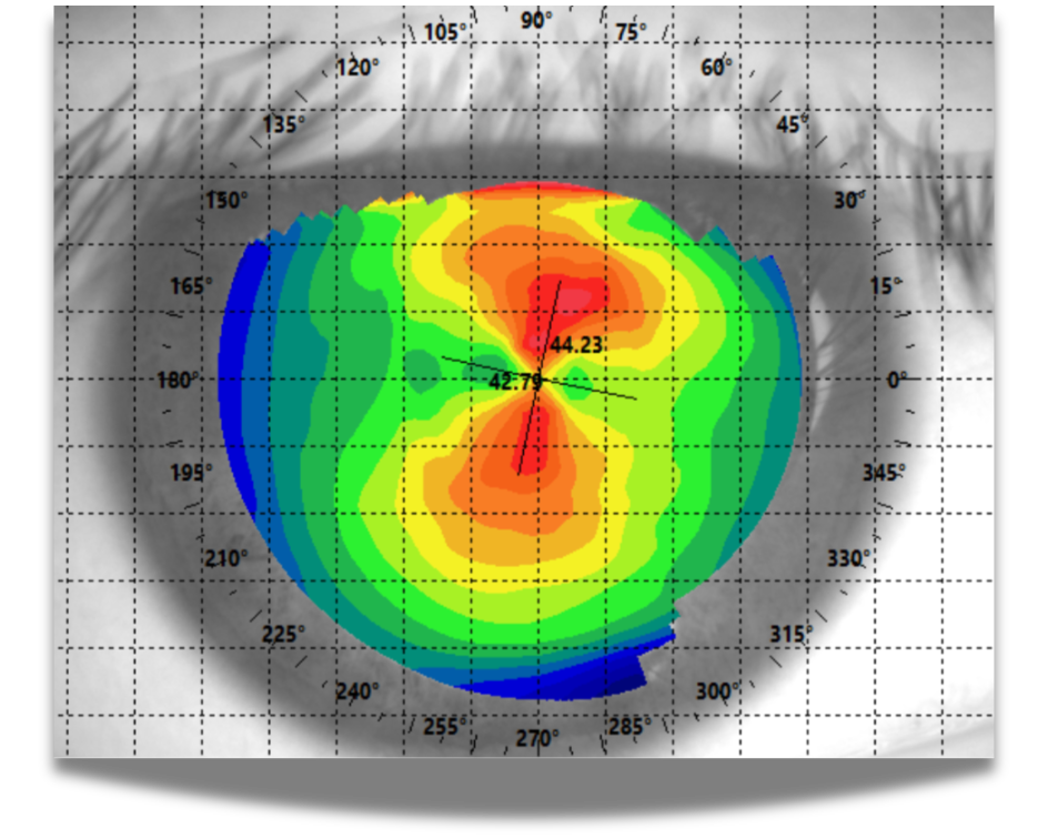
Corneal Topography showing Regular Astigmatism
Treatment and Management
Effectively managing this condition requires specialized treatment which ranges from vision correction to surgical intervention.
- Do not rub your eyes: With keratoconus, try to avoid rubbing your eyes. This can damage thin corneal tissue and make your symptoms worse. If you have itchy eyes that cause you to rub, speak to us about medications to control those symptoms.
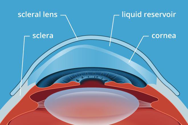
- Specialized Scleral Contact Lenses Traditional glasses or soft contacts often cannot fully correct the vision distortion caused by keratoconus. Instead, patients are frequently fit with rigid, gas-permeable contacts called scleral lenses. Unlike standard lenses, scleral lenses rest comfortably on the white part of the eye (the sclera) and completely vault over the irregular cornea. They create a smooth, fluid-filled artificial surface that allows light to pass through clearly, while still letting essential oxygen reach your eye. Learn MORE about scleral lenses here.
- Cross-Linking: If your condition is actively changing, Dr. Pulis will co-manage your care and refer you to an ophthalmologist for corneal cross-linking. This treatment uses UV light and specialized drops to bond and strengthen the cornea’s collagen fibers, preserving your current vision.